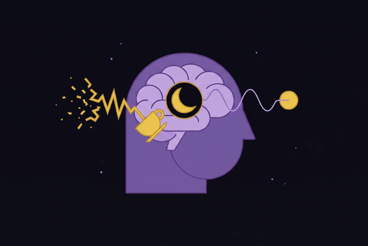
Sleep Stages and Glymphatic Clearance of Brain Waste
Deep sleep, not just hours slept, drives the brain's waste removal.
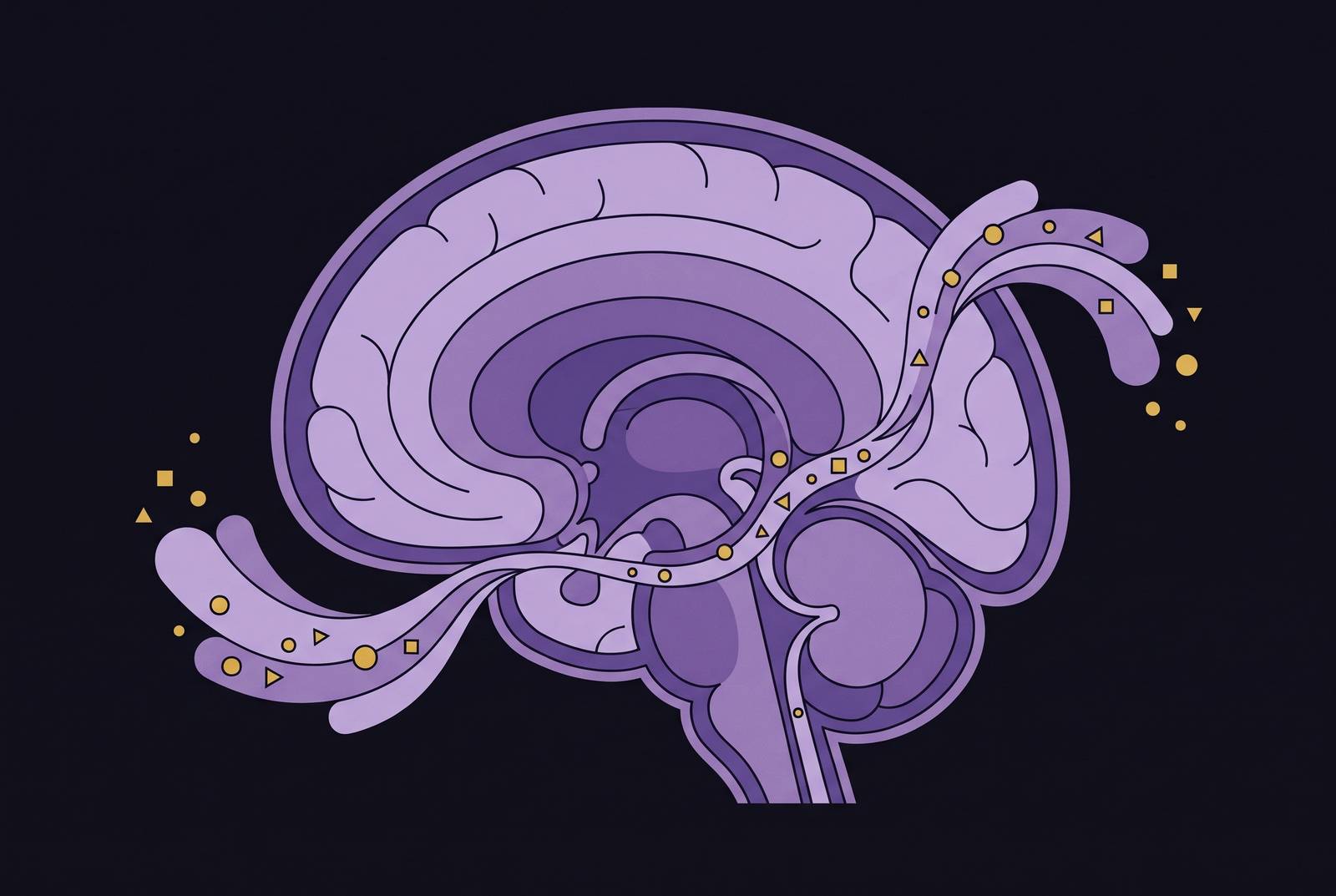
Deep, slow-wave NREM sleep provides a key window for the glymphatic system to remove byproducts such as amyloid-beta and tau. The practical conclusion is plain. Total sleep time is not enough to judge whether sleep is doing its job. The quality and structure of that sleep deserve attention too.
Why the brain needs a dedicated waste clearance system that the rest of the body lacks
An active brain produces metabolic byproducts that must be removed. If they accumulate, some can become harmful. These include amyloid-beta, tau, and α-synuclein, proteins whose buildup is linked to the pathology of Alzheimer’s disease and Parkinson’s disease. The brain therefore faces a basic housekeeping problem. It needs a way to move waste out of the tissue where that waste forms.
Elsewhere in the body, lymphatic vessels help clear extracellular proteins. Brain tissue itself has no such internal vessels, so waste cannot leave by the same route. Cerebrospinal fluid, or CSF, serves a parallel role. Jeffrey Iliff, PhD, at the University of Washington School of Medicine describes CSF as “the 'sink' by which extracellular solutes are 'washed away.'”
The existence of a structured clearance pathway was contested when it was first described in 2012. Juan Piantino’s proof-of-principle contrast MRI study in five human patients visualized fluid moving along defined routes around blood vessels. Clearance depends not simply on having fluid available, but on moving it through a network. This system can falter when the forces that move the fluid grow weaker.
How the glymphatic system actually moves waste out of the brain
Blood vessels provide the scaffold for the glymphatic network. CSF enters along penetrating arteries through spaces bordered by the endfeet of astrocytes, cells that surround the vessels. These routes are called perivascular spaces. From there, CSF exchanges with the fluid between brain cells and picks up dissolved waste. The resulting fluid moves toward routes around veins and onward into downstream drainage pathways.
The plumbing depends on both channels and pumps. Aquaporin-4, or AQP4, is a water-channel protein abundant in astrocyte endfeet that helps regulate this fluid exchange. Arterial pulsations help move the fluid, as do slower changes in vessel diameter. The network feeds into meningeal and cervical lymphatic drainage. Its connection to the meningeal lymphatic system is established, although the full anatomical route has not yet been mapped.
Hauglund and colleagues’ 2025 Cell study identified an upstream driver of this pumping action. A brain region called the locus coeruleus generates oscillations in noradrenaline, which produce rhythmic vessel contractions known as vasomotion. Those contractions physically pump CSF into brain tissue. Sleep position may also influence flow, with evidence favoring side sleeping over sleeping on the back or stomach.
Why glymphatic clearance is gated almost entirely to slow-wave NREM sleep
A night’s sleep usually has four to six roughly 90-minute cycles that move through N1, N2, N3, and REM sleep. N3 is deep, slow-wave NREM sleep, and it is concentrated in the earlier cycles before shrinking toward morning. Glymphatic activity is much greater in this stage than during lighter sleep, REM, or wakefulness. As a result, the timing and composition of sleep affect what a given number of hours can accomplish.
During N3, slow-wave delta activity accompanies an enlargement of the space outside brain cells, opening the routes through which CSF exchanges with tissue fluid. The physical conditions for clearance become more favorable. Glymphatic activity rises with greater delta activity and falls with higher beta activity and heart rate. Losing or fragmenting N3 can therefore disrupt the clearance window even when total sleep time looks adequate. Which part of the night is disrupted also counts, because deep sleep is not evenly distributed.
A 2026 Nature Communications study found causal evidence that physiological processes at work during sleep, especially lower resistance within brain tissue, help clear more Alzheimer’s-associated biomarkers into plasma overnight. A selective suppression experiment approached the question from the opposite direction. Deliberately reducing slow-wave activity raised CSF amyloid-beta levels the following morning.
Even so, the amount of slow-wave activity is not the whole story. A 2026 Scientific Reports study of 47 older adults, with a mean age of 70.5, found that slow wave–spindle coupling was the best predictor of baseline amyloid-beta levels. This is the coordination of slow oscillations with thalamic sleep spindles at 12–15 Hz. It was better at predicting those levels than slow-wave activity alone, age, or cognitive functioning. N3 also supports memory consolidation through coordinated communication between the hippocampus and neocortex, so its internal timing serves more than waste clearance.
What happens to cognition and long-term brain health when slow-wave sleep is chronically disrupted
Disrupted sleep impairs short-term performance. Research from 2025 documented poorer task-switching accuracy after total sleep deprivation, as well as slower reaction time and prolonged auditory P300 latency, a brain-response measure. These findings concern sleep loss more broadly. These findings show why next-day functioning belongs alongside distant disease risk in this discussion, while leaving the contribution of glymphatic failure to the deficits unresolved.
Poor sleep can also become self-reinforcing. It elevates cortisol, while elevated cortisol further degrades sleep quality and memory encoding. The resulting loop can impair executive function during the day and make restorative sleep harder to sustain at night. Over time, poor clearance makes it easier for harmful proteins to build up. Amyloid-beta and tau are linked to Alzheimer’s pathology, while α-synuclein is linked to Parkinson’s pathology.
Those links describe risk and mechanism. Abnormal glymphatic function is also associated with pathological processes in vascular dementia, broadening the concern beyond one protein or diagnosis. Carvalho and colleagues’ 2024 Mayo Clinic Study of Aging examined NREM slow-wave activity in relation to longitudinal amyloid accumulation in older adults with obstructive sleep apnea, or OSA. Repeatedly disrupted sleep is not merely an experimental tool but a real condition in which sleep architecture and protein accumulation can be studied together.
Why the standard fix — sedative sleep aids — may make glymphatic clearance worse
The counterintuitive turn is that producing more sleep may not preserve the machinery that makes sleep restorative. A sedative can increase time asleep without reproducing every feature of natural N3. That distinction is especially important for the noradrenaline oscillations that drive vasomotion. Quieting brain activity and sustaining a fluid pump are not necessarily the same biological task.
The Hauglund study found that zolpidem impaired noradrenaline-driven oscillations and reduced glymphatic inflow in mice. The drug acts by enhancing GABAergic signaling. In that experiment, sedation disrupted part of the mechanism needed to move fluid through brain tissue. This is a mechanistic warning whose implications for long-term harm in people remain unproven. It also does not establish that every sleep medication has an identical effect.
The distinction becomes particularly relevant when disrupted sleep already has an underlying cause, as in OSA. More sedated hours cannot by themselves show that slow-wave activity or clearance has improved. Sedative sleep aids can still have valid uses. Sedative sleep aids should be evaluated by more than the duration of sleep they produce, and this animal finding should not be treated as a reason to reject a particular medication.
What the science currently supports for improving slow-wave sleep quality without drugs
Non-drug approaches work at different points in the process. Some try to strengthen slow waves during sleep, while others support the conditions that allow good sleep architecture to develop. These approaches complement one another and depend on sleep. Their evidence also differs. Boosting a sleep signal, reducing cortisol, and demonstrating better waste clearance are separate outcomes.
Closed-loop auditory stimulation, or CLAS, targets slow-wave activity directly. It delivers sounds timed to a particular phase of the slow oscillation during N3, and multiple studies have shown that it can boost slow waves. A clinical trial is assessing whether repeated nights of CLAS can also enhance glymphatic clearance. That second step remains a research question.
The completed SIESTA Trial, registered as NCT03954210, was designed to test sleep intervention as a way to enhance cognitive status and reduce beta-amyloid. Completion alone does not establish a successful outcome.
Upstream approaches focus partly on the stress response. Chronically elevated cortisol can fragment sleep and suppress N3, making the hypothalamic-pituitary-adrenal axis, or HPA axis, relevant to sleep quality. Ashwagandha’s withanolides affect this system and GABAergic signaling, with anti-inflammatory and antioxidant actions also described. A 2012 randomized, double-blind, placebo-controlled trial in the Indian Journal of Psychological Medicine found that 300 mg of full-spectrum root extract twice daily reduced serum cortisol during the trial. Further trials have reported improvements in sleep quality, latency, efficiency, and total sleep time, but these outcomes do not establish enhanced glymphatic clearance.
Rhodiola rosea is another proposed upstream support. It appears to modulate the HPA axis, support healthy serotonin and dopamine activity, and reduce stress-related cortisol spikes during mental or physical overload. That makes it relevant to chronic mental fatigue and burnout contexts.
Lion’s Mane contains hericenones and erinacines that stimulate nerve growth factor production, a mechanism relevant to neuronal support. Human results need a narrower reading. A 2025 randomized controlled trial in healthy younger adults found no significant overall improvement in cognition or mood after a single dose of standardized extract, apart from an isolated pegboard-test improvement. Bacopa monnieri has stronger repeated-dose findings for some cognitive outcomes. Multiple double-blind, placebo-controlled trials reported improvements in delayed recall, information processing speed, and working memory after 8–12 weeks, while its bacosides may also support BDNF, a protein involved in neural resilience.
None of those ingredient findings demonstrates a glymphatic treatment. Absorption also limits what an ingredient can do, so delivery format and bioavailability require assessment beyond a label. For everyday sleep support, the recommended habits are more basic. Consistent sleep timing and light management support circadian alignment and protect opportunities for N3. Irregular schedules can compress those opportunities even when the nightly hour count appears sufficient.
What honest caveats remain — and why they strengthen rather than weaken the case for prioritizing sleep stage quality
There is no validated, non-invasive bedside or wearable measure of glymphatic flow. Current research tools include diffusion MRI measures such as the ALPS index, enlarged perivascular spaces on T2 MRI, and intrathecal contrast studies. Each has limits. ALPS takes samples from white matter near the lateral ventricles rather than gray matter, and enlarged perivascular spaces can arise from several biological processes. These are not practical consumer measures of overnight brain cleaning.
The full chain from a sleep intervention to improved clearance to lasting cognitive protection in living humans is inferred partly from biomarkers and awaits direct demonstration from end to end. Experimental findings support causal steps within that chain. They do not make every proposed intervention effective.
The underlying model also has unresolved details. A 2017 paper challenged AQP4’s role after finding solute transport that was diffusive and AQP4-independent. Iliff attributes the discrepancy to differences in experimental methods. Findings from different imaging methods, species, and research groups back the core clearance pathway. Its exact mechanics remain open to refinement.
These limits make precision more useful than certainty. Better-structured N3 sleep is linked across research lines to more favorable amyloid-beta biomarkers and cognitive outcomes, while intervention studies are testing how far those benefits can be changed deliberately. The goal is not simply more hours unconscious, but sleep with the structure the brain needs.
Sources
- Clearance mechanisms of the glymphatic/lymphatic system in the brain: new therapeutic perspectives for cognitive impairment
- When sleep fails, brain clearance suffers: the role of glymphatic impairment in clinical neurology
- Norepinephrine-mediated slow vasomotion drives glymphatic clearance during sleep: Cell
- The Brain’s Waste Disposal System and Why Sleep Is Key for Brain Health
- Glymphatic system in neurological disorders and implications for brain health
- The glymphatic system clears amyloid beta and tau from brain to plasma in humans
- Deep sleep slow wave–spindle coupling is selectively linked to plasma amyloid-β levels in older adults in clinical trials
- Sleep reduces CSF concentrations of beta-amyloid and tau: a randomized crossover study in healthy adults - PMC